

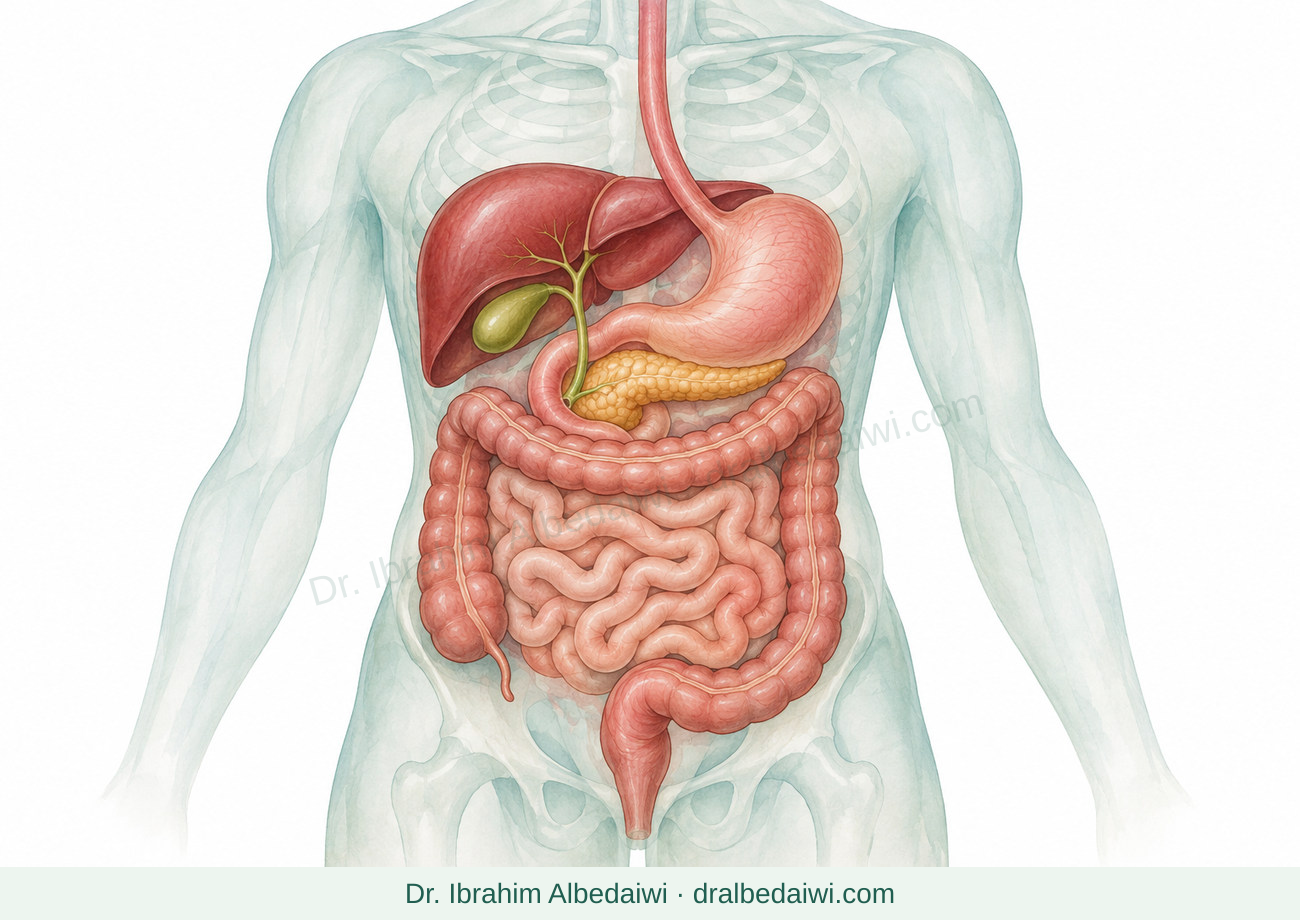

Benign liver lesions and cysts
Content date: 10 November 2026
A liver lesion does not automatically mean cancer. A confirmed simple cyst, hemangioma or FNH is usually reassuring, while a hepatic adenoma needs a different plan.
Your next step
Bring your images and medicine list. Ask: Is this a simple cyst, hemangioma, FNH or adenoma, and what follow-up is needed?
Get urgent help: Sudden severe upper abdominal pain needs urgent assessment even without dizziness. Fainting, pallor or sweating with pain, or fever with marked illness, requires emergency care.
What does a liver lesion or mass mean?
- A lesion is an area that looks different on a scan, often found incidentally. It may contain fluid, blood vessels or liver cells. “Lesion” is a description, not a diagnosis; “tumor” does not necessarily mean cancer.
- Assessment considers images, symptoms, previous liver disease or cancer, and medicines. Not every pain comes from the lesion, and size alone does not determine treatment.
How is the type identified?
- Ultrasound may suffice; otherwise, liver-specific multiphase contrast MRI or CT may be needed. Specialized MRI contrast helps distinguish FNH from adenoma. Report possible pregnancy, kidney disease or previous contrast reactions.
- Biopsy, or tissue sampling, is reserved for selected uncertain findings after specialist review. Biopsy of a suspected hemangioma is avoided when possible because it can bleed.
Simple cyst: a fluid-filled pocket
- A confirmed simple cyst has a thin wall and characteristic scan appearance. It is not cancer and does not become cancer. Without symptoms, treatment or regular scans are usually unnecessary.
- If it causes confirmed pressure-related symptoms, drainage with treatment of the cyst wall or surgery may help. Drainage alone can allow refilling. Internal partitions, nodules or an irregular wall need further assessment; not every cyst is simple.
Hemangioma: a cluster of blood vessels
- This benign blood-vessel cluster is not cancer or a precancerous lesion. It is sometimes informally called a “blood cyst” in Arabic, but it is not a simple fluid-filled cyst. Its contrast-filling pattern often establishes the diagnosis; ultrasound may suffice in typical cases.
- A secure diagnosis without symptoms or special risks usually needs neither treatment nor repeated scans. Size alone does not require surgery. Bleeding or significant pressure on nearby organs may prompt intervention; complications are rare.
Focal nodular hyperplasia (FNH)
- FNH is localized benign liver-cell growth, thought to respond to altered blood flow. It is neither cancer nor a precancerous lesion.
- Imaging-confirmed FNH usually needs neither treatment nor regular surveillance. Atypical imaging or symptoms prompt specialist review of the diagnosis and other possible causes before surgery is considered.
Hepatic adenoma: a different type of benign tumor
- An adenoma arises from liver cells and differs from FNH. Estrogen, anabolic steroids and excess weight may be associated with it. Some adenomas bleed or rarely become cancer, so assessment and follow-up matter even without symptoms.
- Subtype, size, growth and sex guide the plan. Some cases can be monitored; intervention is more likely in men, growing or large lesions, or high-risk subtypes. Not every adenoma needs immediate surgery.
- The team reviews hormones, contraception and anabolic steroids, and recommends weight loss when appropriate. Arrange suitable alternative contraception before changes. Selected treatments include removal, blocking feeding blood vessels by embolization, or ablation.
Pregnancy and contraception: the lesion type matters
- Confirmed FNH or hemangioma usually does not prevent pregnancy or automatically require stopping contraception. Large, symptomatic or uncertain lesions need individual review.
- With adenoma, discuss pregnancy beforehand with liver and maternity teams: growth may require closer monitoring or treatment before conception. Size and features guide the plan; FNH advice does not apply.
Are all cysts and lesions alike?
- Complex or parasitic (hydatid) cysts and polycystic liver disease differ from simple cysts. Travel, animal exposure and family history may matter. Uncertain findings need assessment; a suspected parasitic cyst must not be sampled outside a specialist plan.
Make follow-up clear
- Bring old and new images, reports and a medicine list. Ask for the diagnosis, how certain it is, and whether the next scan monitors the lesion or the liver overall.
- A confirmed benign lesion does not cancel liver cancer surveillance needed for F4 cirrhosis or hepatitis B. Your team determines the plan for the underlying liver disease separately from the lesion.
Quick comparison after diagnosis is confirmed
| Type | What is it? | Usual plan without symptoms or special circumstances |
|---|---|---|
| Simple cyst | Fluid-filled pocket | Usually no treatment or routine scans. |
| Hemangioma | Cluster of blood vessels | Usually no intervention once confirmed; consider background liver risks. |
| FNH | Localized benign growth of liver cells | Usually no treatment or routine surveillance. |
| Adenoma | Liver-cell tumor that can bleed or change | Specialist follow-up; hormone changes or intervention may be needed. |
This comparison excludes uncertain lesions and complex cysts. It does not replace monitoring of underlying liver disease.
© 2026 Dr. Ibrahim AlbedaiwiWhen to get help
Sudden severe upper abdominal pain needs urgent assessment even without dizziness. Fainting, pallor or sweating with pain, or fever with marked illness, requires emergency care.
Persistent pain or fullness, unexplained weight loss, jaundice, or a report requesting further testing: arrange review. Contact the team early when planning pregnancy with an adenoma.
Related guides
Liver cancer: diagnosis and treatment